








Proper Care of Hardworking Leather

Sno-Seal provides maximum waterproofness and durability for smooth full-grain leather footwear. Oil or chrome tanned leather accepts Sno-Seal readily after removal of any factory coating. (Glycerin or Carnuba, wax which are water-soluble, is often applied for a temporary shine and scuff resistance). A new boot should be washed with a soft brush using Sport-Wash or a mild liquid dish washing soap, and allowed to dry completely before applying Sno-Seal.

Sno-Seal also protects split, sanded, suede, rough and recycled leather. These materials need the protection of Sno-Seal for rugged use, but application of Sno-Seal will change their appearance. As Sno-Seal coats the exposed fibers and fill surface gaps, these non-top grain materials become darker and slicker.

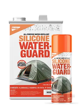
Sno-Seal can also be used on silicone treated leather when the silicone is scuffed and worn off and no longer works. Spray or dauber application of ATSKO’s Silicon Water Guard. is quick, easy, and superior to any other silicone, but no silicone can provide the durable waterproofness of Sno-Seal. When silicone treated boots can no longer resist water, simply scrub the remaining silicone out of the leather with Sport-Wash, saddle soap or dish detergent. Then, slowly dry them for a couple of days and apply Sno-Seal according to directions.

Gore-Tex® fabric lined boots should be protected with Sno-Seal the same as you would treat them if Gore-Tex® was not present. Sno-Seal does not effect Gore-Tex® and Gore-Tex® does nothing to protect the outer boot form water and other abuse. To benefit from the breathability of the Gore-Tex® liners, the inside of the leather must be dry and free of oil, grease, or animal fat so that it can absorb perspiration from the foot. At night this moisture will evaporate thru the Gore-Tex® and pass out of the boot leaving it dry and comfortable for the next day’s use. The Gore-Tex® line will hold water away from your foot, but Sno-Seal will keep your boot warmer and lighter by preventing water from being absorbed by the outer layer of the boot or leaking into the space between the leather and the liner. When the leather is wet it can add 1 1/2 pounds to a boot.

Why is Sno-Seal so good?
The superiority of Sno-Seal results from the combination of its ability to remain fixed in the outer layer of the leather and the superior water resistance of beeswax. Beeswax has a melt point of about 146º F. It is very dry and oil free. Thus once applied it is not only waterproof but extremely durable.

Sno-Seal provides just enough lubrication to prevent hardening of leather in typical use. It does not soften leather. It was invented in 1933 by Ome Daiber to waterproof leather mountain climbing and downhill ski boots without destroying the stiffness required to climb or steer a ski. Boots, gloves, holsters etc. will maintain their strength for years if treated only with Sno-Seal. A baseball mitt can be softened with a light application of mineral oil. When exactly the right ball pocket and fit have been achieved, applying Sno-Seal will maintain exactly the same form for the life of the glove.

Grease, oil, animal fat, and most other wax formulas are liquid (or at least soft enough to migrate thru leather) at wearing and storage temperatures. Sno-Seal is not. After application, when the volatile components have done their task of drawing Sno-Seal into the warmed leather, they evaporate completely leaving solid wax that cannot migrate at less than 146° F.

Similar methods of waterproofing
Grease, animal fat and oil, including some brands of silicone oil, soak away from the surface and penetrate through the leather. They require frequent re-applications to maintain effective concentration on the surface. After a time the heel counters and box toes soften. The leather becomes soft and spongy then the product begins to appear on your socks indicating it has soaked all thru the leather. Animal fats become rancid, decaying leather and promoting fungal growth until one day the leather is so rotten the eyelets pull out when you lace up your boots. Saturated leather cannot absorb perspiration and loses its insulation value so your foot feels clammy and cold. With Sno-Seal, the beeswax stays where you need it, in the outer surface of the leather. The balance of the thickness remains open and dry for insulation and the absorption of foot moisture. By resisting migration Sno-Seal assures that there will be no weakening of thermal or adhesive bonds, a common problem with silicones and other liquids.

Who can you trust when choosing a waterproofing product?
For obvious reasons, many Boot marketers recommend a waterproofing product with their name on it, even if something else is better. Perhaps the surest way to get the right product for a boot is to find a Boot manufacturer that provides a sample of a waterproofing product (one they don’t make themselves) in each pair of new boots. Boot manufacturers experience a dramatic reduction in warrantee claims when they encourage customers to properly care for leather boots by providing a compatible product. From L.L. Bean to Sorel, more new boots, around the world, include a sample of Sno-Seal than of any other brand.

Other uses of Sno-Seal include gloves, oiled canvas coats, chaps, belts, hats, and any other wearable leather that is exposed to water. Topsiders and other leather footwear and gloves can easily last 4 times as long in severe salt water use if protected with Sno-Seal.

Industrial environments including acid, alkaline, blood, and abrasives can shorten the life of shoes, boots, gloves, aprons etc. These leather items will last longer, clean up easier, and provide better protection if maintained with Sno-Seal.

Wood can be wax finished with Sno-Seal for a durable “unfinished” look. Sno-Seal can be applied over an oil stain for a satin finish that resists water damage.

Equestrians recognize the value of Sno-Seal for harnesses, saddles, scabbards and all leather protection. Sno-Seal will make an old horse blanket absolutely waterproof. Farriers apply Sno-Seal to hoofs to prevent softening from excess moisture and also to prevent brittleness and splitting from excessive dryness.

For over 60 years customers have been telling us about their great results using Sno-Seal to protect severely dry abused skin, hands, feet, knees, elbows, lips, etc. A new formulation of Sno-Seal has been created for these applications. Pro-Tech-Skin,, a similar beeswax formula, is now available for application to the skin. Pro-Tech-Skin, has already proven itself effective for psoriasis, diaper rash, and dry and cracking skin and cuticles associated with frequent washing, dry cold weather, and advancing age. By sealing in natural moisture Pro-Tech-Skin, allows your skin to heal itself.

Silicones offer easy application but less water resistance and protection than Sno-Seal.
Silicones resist water 2 different ways. The first is similar to wax, oil, animal fat, and grease: it simply occupies space in the leather so that water can’t occupy the same space. This is of limited value compared to Sno-Seal and other wax products that occupy space better, more permanently and with less migration. The second method is by reducing surface tension so that water beads up and does not “wet” it. This is similar to the function of Fluoropolymers but Fluoropolymers – when successful – reduce surface tension further so that even oil and some solvents can’t “wet” it. (This is why Fluoropolymer is superior for stain resistance.)

Silicones are usually in solvent systems for water repellent products because water systems (aqueous emulsions of silicone) can’t match the performance of the solvent systems. A variety of solvents will work but there are advantages and disadvantages with each.

ATSKO Inc. makes a Heavy Duty catalyzed silicone product called Silicon Water Guard
The only silicone product to score 100 on the ASTM D1913 water spray test. We use a premium solvent that has a higher flash point, (less dangerous because it will not flash until it reaches a much higher temperature), lower odor and the most environmental acceptance of any in its category. This solvent requires more drying time but we believe that our customers would make the same choice if given the facts. Because we use a high concentration of top quality catalyzed silicone (a resin that cross-links for maximum durability), Water-Guard delivers effective repellence with a single application saving the time, cost, and solvent of a second application. Silicon Water Guard comes in an all steel can that can be recycled. Steel is the most recycled material in the world. Our Aerosol propellants is harmless C02. It is collected as a byproduct of other processes and only two grams Of C02 are necessary to empty a 12 ounce can. Many of our competitors use over 2 1/2 ounces of explosive Propane-Isobutane gas as propellant. Using fuel as a propellant does not improve their products but it is very heavy, very cheap, and is included in their statement of net weight.

Fluoropolymers are designed and intended for use on textiles for stain protection. They require an absolutely clean surface for adherence and 300° F heat is used at the mill to orient the molecules for maximum effectiveness. Applied under perfect conditions they work well and last for many washings if washed only in Sport-Wash residue free detergent.

Fluoropolymers are generally superior for oil and stain repellency but generally inferior to silicone and waxes for water repellency. Factory applied Fluoropolymer treatments on textiles can be restored to full effectiveness by washing with Sport-Wash to remove residue and then Ironing on Steam setting.

Fluoropolymers can give inconsistent results depending greatly upon what surface they are applied to. Baking at 300° F as in textile mill applications is impossible for boots and it is also difficult to keep boots clean enough for maximum repellency. In any case Fluoropolymers are better left for applications where Appearance is more important than Long Term Heavy Duty Waterproofness.

What is the bottom line for boot protection?
When appearance is more important than waterproofness, especially on light suede, consider Fluoropolymers like Scotchguard or Teflons. On cloth shoes you can use our water based Permanent Water-Guard. Use Silicon Water Guard for silicone tanned, and rough finished leather. Use Sno-Seal for oil and chrome tanned full grain leather. Sno-Seal has been the most effective, longest lasting, and best protection for hard working leather since 1933. Sno-Seal the original Beeswax Waterproofing is still the best today. Sno-Seal is now available in Black and Brown.

Leather Care for Ice Skates

The comfort of real leather will never be matched because of its combination of strength, support, breathability, absorbency, elasticity, warmth, and feel. Proper care will maintain these features and extend the useful life of the skate. The upper or shoe must provide correct support to maximize performance and reduce injuries. Water proofing products like grease, oil, and animal fat soak through and weaken the leather, reducing control and the support needed for maximum performance. As a skating shoe is softened by conventional waterproofing it changes in feel and response. Skating technique must be modified to overcome the weakness of the deteriorating shoe. This slows learning and destroys consistency.

Sno-Seal®, the original beeswax waterproofing, was invented in 1933 to protect ski boots and ice skates without softening or weakening them. It maintains the strength and shape of the boot for consistent edge control and the ability to transfer weight fore and aft. It has just enough solvent to allow easy application but this evaporates as it dries to a solid wax. Sno-Seal® remains in the outer surface of the leather, where it is needed to keep the shoe dry. It preserves the life of the leather and lasts a lot longer than liquid products because it stays put.

Always dry blades, blade supports, soles, and uppers immediately after skating. Interior drying does not begin until the skate reaches room temperature and is greatly delayed if ice and water left on the outside cool the inside to below room temp. Skates should not be stored in a closed container until completely dry. The entire process takes a lot longer if any water penetrates the leather from the outside so waterproofing is very important. Sno-Seal® is all wax so it is compatible with shoe polish. Sno-Seal® should be applied first, and any excess buffed off before applying a polish for appearance.

Sno-Seal®, the original beeswax waterproofing, cannot migrate through the leather. It remains concentrated in the outer surface of the leather and does not wick in. The inside of the leather remains open and porous enabling it to breathe freely, removing excess moisture from the foot. This provides hours of comfort during skating and when the skate is removed any remaining moisture evaporates so the skate will soon be ready for another session.

Sno-Seal® is also critical on the bottom or sole of the skate. Custom skate builders apply Sno-Seal® in the screw holes and between the blade mounts and sole to prevent water from entering. Water in the screw holes will rust some screws and it will always swell and weaken the leather. A water swelled sole changes the alignment between the blade and the shoe. Sno-Seal® should be reapplied to the sole, especially around the blade mounts, every time you treat the upper, sole edges, or welt stitching.

UV Color and Vision References

Popular Publications

  1. CLANCY, UV The Invisible Factor. Petersen’s Bowhunting. August 1990 pp 46-50.
  2. GOOCH, BOB. Blaze in the Woods. OUTDOOR LIFE November 1992 pp 90-102.
  3. MANDILE, TONY. Now they see you…Now they don’t. OUTDOOR LIFE July 1990 pp 81-90.
  4. Charles J. Alsheimer The Role of Color Vision in Whitetails. Deer & Deer Hunting December 1999 pp 20-28
  5. ROBB, BOB The Question of COLOR. Whitetail Journal October 2000 pp 53-56
  6. The Behavioral Test to Examine the Evolution of Color Vision in Vertebrates Link
  7. https://www.vri.cz/docs/vetmed/54-8-351.pdf
  8. How Birds Keep Secrets in Color, By Nick Atkinson([email protected]) | April 27, 2005

Books

  1. JACOBS, GERALD H., The Distribution and Nature of Colour Vision among the Mammals Department of Psychology, University of California, Santa Barnara 1 March 1993.
  2. LYTHGOE, J. N., The Ecology Of Vision. Clarendon Press, Oxford, 1979.
  3. RODIECK, R. W., The Vertebrate Retina Principles Of Structure And Function. W. H. FREEMAN & CO.
  4. SINCLAIR, S., Foreword by DEAN YEAGER, PHD., How Animals See, Other Visions of Our World. Facts On File Publications, New York.
  5. WALD, G., Edited by Richard Jung, Life & Light Handbook of Sensory Physiology Volume VII/3 Central Processing of Visual Information. Springer-Verlag, New York, 1973.
  6. IVOSEVIC, S. W., Birder Camouflage on disguising appearance, movements, and location while watching or photographing birds. Stanley W. Ivosevic, Castle Hayne, 2000.

Juried Scientific Publications

  1. ANDERSSON, M. (1996) Sexual Selection (Princeton Univ. Press, Princeton, NJ).
  2. ANDERSSON, S. (1996) Proc. R. Soc. London Ser. B263, 843-848.
  3. AGUIRRE, G .(1978). Retinal degenerations in the dog, I: Rod dysplasia. Experimental Eye Research 26, 233-253.
  4. AGUIRRE, G., ALLIGOOD, J, O’BRIEN, P. & BUYKMIHCI, N. (1982). Pathogenesis of progressive rod-cone degeneration in minature poodles. Investigative Ophthalmology and Visual Science 23, 610-630.
  5. AHNELT, P. K. (1985). Characterization of the color-related receptor mosaic in the ground squirrel retina. Vision Research 25, 1557-1567.
  6. ALEXANDER, G. & STEVENS, G. (1979). Discrimination of colours and gray shades by Merino ewes: tests using coloured lambs. Applied Animal Ethology 5, 215-231.
  7. ALI, M. A. & KLYNES, M. A. (1985). Vision in Vertebrates. Plenum Press, New York.
  8. ALPERN, M., LEE, G. B., MAASEIDVAAG, F. &- MILLER, S. S. (1971). Colour vision in blue-cone monochromacy. Journal of Physiology 212, 211-23 3.
  9. BAILEY, W. J., SLICHTOM, J. I,. & GOODMAN, M. (1992). Rejection of the ‘Flying Primate’ hypothesis by phylogenetic evidence from the y-globin gene. Science 256, 86-89.
  10. BARLOW, H. B. (1982). What causes trichromacy? A theoretical analysis using comb-filtered spectra. Vision Research 22, 63S-643.
  11. BAUCHEMIN M. L: The fine structure of the pig’s retina. Albrecht von Graefes Arch Klin Exp Ophthalmol 190:27-45, 1974.
  12. BAYLOR, D. A., NUNN, B. J. & SCHNAPF, J. I,. (1984). The photocurrent, noise and spectral sensitivity of rods of the monkey. Macaca fascicularis. Journal of Physiology 357, 575-607.
  13. BAYLOR, D. A., NUNN, B. J. & SCHNAPF, J. I,. (1987). Spectral sensitivity of cones of the monkey Macaca fascicularis. Journal Of Physiology 390, 145-160.
  14. BENNETT, A. T. D. & CUTHILL, I. C. (1994) Vision Res.34, 1471-1478
  15. BENNETT, A. T. D., CUTHILL, I. C. & NORRIS, K. J. (1994) Am. Nat.144, 848-860
  16. BENNETT., A. T. D., CUTHILL, I. C., PARTRIDGE, J.C.& MAIER, E. J. (1996) Nature (London)380, 433-435
  17. BENNETT., A. T. D., CUTHILL, I. C., PARTRIDGE, LUNAU, K. (1997) Ultraviolet plumage colors predict mate preferences in starlings. Proc. Natl. Acad. Sci. USA Vol 94,,pp. 8618-8621
  18. BIRCH, D. & JACOBS, G. H. (1975). Behavioral measurements of rat spectral sensitivity. Vision Research 15, 687-691.
  19. BLACKWELL, H. R. & BLACKWELL, 0. M. (1961). Rod and cone receptor mechanisms in typical and atypical congenital achromatopsia. Vision Research 1, 62-107.
  20. BLAKESLEE, B. & JACOBS, G. H. (1982). Color vision in the spider monkey (Ateles). Folia Primatologica 38, 86-98.
  21. BLAKESLEE, B. & JACOBS, G. H. (1985). Color vision in the ring-tailed lemur (Lemur catta). Brain, Behavior and Evolution 26, 154-166.
  22. BLAKESLEE, B., JACOBS, G. H. & NEITZ, J. (1988). Spectral mechanisms in the tree squirrel retina. Journal of Comparative Physiology A 162, 773-780.
  23. BOWMAKER, J. K. Trends Neurosci., 196, 1980.
  24. BOWMAKER, J. K. (1984). Microspectrophotometry of vertebrate photoreceptors. Vision Research 24, 1641-1650.
  25. BOWMAKER, J. K. (I 991a). Visual pigments, oil droplets and photopigments. In The Perception of Colour (ed. P. Gouras), pp. 108-127. CRC Press, Boca Raton.
  26. BOWMAKER, J. K. (1991b). Visual pigments and colour vision. In From Pigments to Perception : Advances in Understanding Visual Processes (ed. A. Valberg and B. B. Lee), pp. 1-9. Plenum Press, New York.
  27. BOWMAKER, J. K. (1991c). The evolution of vertebrate Visual pigments and photoreceptors. In Evolution of the Eye and Visual System led. J. R. Cronly-Dillon and R, L. Gregory), pp. 63-81. CRC Press, Boca Raton.
  28. BOWMAKER, J. K., ASTELL, S., HUNT, D. M. & MOLLON, J. D. (1991). Photosensitive and photostabile pigments in the retinae of Old World monkeys. Journal of Experimental Biology 156, 1-19.
  29. BOWMAKER, J. K., DARTNALL, J. A., LYTHGOE, J. N. & MOLLON, J. D. (1970). The visual pigments of rods and cones in the rhesus monkey, Macaca mulatta. Journal of Physiology 274, 329-348.
  30. BOWMAKER, J. K., DARTNALL, H. J. A. & MOLLON, J. D. (1980). Microspectrophotometric determination of four classes of photoreceptor in an Old World primate, Macaca fascicularis. Journal of Physiology 298, 131-143.
  31. BOWMAKER, J. K., JACOBS, G. H. & MOLLON, J. D. (1987). Polymorphism of photopigments in the squirrel monkey: A sixth phenotype. Proceedings of the Royal Society of London B231, 383-390.
  32. BOWMAKER, J. K., JACOBS, G. H., SPIEGELHALTER, D. J. & MOLLON, J. D. (1985). Two types of trichromatic squirrel monkey share a pigment in the red-green spectral region. Vision Research 25, 1937-1946.
  33. BOYNTON, R. M. ‘Human Color Vision’. Holt-Rinehart-Winston, New York, 1979.
  34. BRINDLEY, G. S. (1970). Physiology of the Retina and Visual Pathway.), 2nd edn. Williams & Wilkins, Baltimore.
  35. BURKHARDT, D. (1982). Birds, berries and UV. Naturwissenschaften 69, 153-157.
  36. BURKHARDT, D. (1983). Wavelength perception and colour vision. Symposia of the Society for Experimental Biology 36, 371-397.
  37. BURKHARDT, D. & FINGER, E. (1991). Black, white, and UV: how birds see birds. Naturwissenschaften 78, 279-280.
  38. BURN, D. M. (ed.) (1991). The Encyclopedia of the Animal World. Peerage Books, London.
  39. BUSCH, H. & DUCKER, G. (1987). Das visuelle Leistungsvermogen der Seebaren (Arctocephalus pusillus und Arctocephalus australis). Zoologischer Anzeiger 219, 197-224.
  40. CAMPENHAUSEN, C. von. (1986). Photoreceptors, lightness constancy and color vision. Naturwissenschaften 73, 674-675.
  41. CHAUSSEIL, M. (1992). Evidence for color vision in procyonides: Comparison between diunal coatis (Nasua) and nocturnal kinkajous (Potos flavus). Animal Learning and Behavior 20, 259-265.
  42. CICERONE, C. M. (1976). Cones survive rods in the light-damaged eye of the albino rat. Science 194, 1183-1185.
  43. CICERONE, C. M. & NERGER, J. L (1980). The relative numbers of long-wavelength-sensitive to middle-wavelength-sensitive cones in the human fovea centralism Vision Research 29, 115-128.
  44. COCKAYNE, E. A. (1924). The Distribution of Fluorescent Pigments in the Lepidoptera. Trans. Entom. Soc. London, 1924,pp. 1-19.
  45. COILE, D. C. (1982). Adetermination of critical flicker fusion as a function of light intensity in dogs using conditioned supression. Unpublished Masters Thesis, Florida State Univ.
  46. COHEN, A. I. (1972). Rods and cones. In Physiology of Photoreceptor Organs (ed. M. G. F. Fuortes), pp. 63-110. Springer Verlag, Berlin.
  47. CORBET, G. H. & HILL, J. E. (1991). A World List of Mammalian Species, 3rd edn. Oxford University Press, Oxford.
  48. CRESCITELLI, F. (1963). Obituary: Gordon Lynn Walls. Vision Research 3, 1-7.
  49. CRESCITELLI, F. & POLLOCK, J. D. (1972). Dichromacy in the antelope ground squirrel. Vision Research 12, 1553-1586.
  50. CROCKER, R. A., RINGO, J., WOLBARSHT, M. L. & WAGNER, H. G. (1980). Cone contributions to cat retinal ganglion cells receptive fields. Journal of General Physiology 76, 763-768.
  51. CRONIN, T. W. & MARSHALL, N. J. (1989). A retina with at least ten spectral types of photoreceptors in a mantis shrimp. Nature 339, 137-140.
  52. CURCIO, C. A., ALLEN, K. A., SLOAN, K. R., LEREA, C. L., HURLEY, J. B., KLOCK, I. B. & MILAM, A. H. (1991). Distribution and morphology of human cone photoreceptors stained with anti-blue opsin. Journal of Comparative Neurology 312, 610-624.
  53. DALRYMPLE, B. W.: When can your quarry see you? Outdoor Life 156:61, 1975.
  54. DARTNALL, H. J. A., ARDEN, G. B., IKEDA, H., LUCK, C. P., ROSENBERG, C. M., PEDLER, H. & TANSLEY, K. (1965). Anatomical, electrophysiological and pigmentary aspects of vision in the bush baby: an interpretative study. Vision Research 5, 399-424.
  55. DARTNALL, H. J. A. & LYTHGOE, J. N. (1965). The spectral clustering of visual pigments. Vision Research 5, 81-100.
  56. DARWIN, C. (1871) The Descent of Man and Selection in Relation to Sex (Murray, London).
  57. DAW, N. W. & PEARLMAN, A. L. (1969). Cat colour vision: one cone process or several. Journal of Physiology 201, 745-764.
  58. DAW, N. W. & PEARLMAN, A. L. (1970). Cat colour vision: Evidence for more than one cone process. Journal of Physiology 211, 125-137.
  59. DAWIS, S. M. (1981). Polynomial expressions of pigment nomograms. Vision Research 21, 1427-1430.
  60. DAWSON, W. W, (1980). The cetacean eye. In Cetacean Behavior: Mechanisms and Functions (ed. L. M. Herman), PP. 53-100. John Wiley, New York.
  61. DEEGAN II, J. F. & JACOBS, G. H. (1993). On the identity of the cone types of the rat retina. Experimental Eye Research 56, 375-377.
  62. DERRINGTON, A. M., KRAUSKOPF, J. & LENNIE, P. (1984). Chromatic mechanisms in lateral geniculate nucleus of macaque. .Journal of Physiology 357, 219-240.
  63. DE VALOIS, R. L. & DE VALOIS, K. K. (1988). Spatial Vision. Oxford University Press, New York.
  64. DE VALOIS, R. L. & JACOBS, G. H. (1984). Neural mechanisms of color vision. In Handbook of physiology. Section r. Volume III. Sensory Processes (ed. I. Darian-Smith), PP. 425-456. Williams & Wilkins, Baltimore.
  65. DE VALOIS, R. L., MORGAN, H. C., POLSON, M. C., MEAD, W. R. & HULL, E. M. (1974). Psychophysical studies of monkey vision. I. Macaque luminosity and colour vision tests. Vision Research 14, 53-67.
  66. DODT, E. (1967). Purkinje shift in the rod eye of the bush baby, Galago crassicaudatus. Vision Research 7, 509-517.
  67. DUBIN, M. W. & TURNER, L. (1977). Anatomy of the retina of the mink (Mustela vison). Journal of Comparativ Neurology T,73, 275-288.
  68. DUCKER, G. (1964). Colour-vision in mammals. Journal of the Bombay Natural History Society 61, 572-586.
  69. EBREY, T. G. & HONIG,B. (1977). New wavelength-dependent visual pigment nomograms. Vision Research 17, 147-151.
  70. EENS, M., PINXTEN, R. & VERHEYEN, R. F. (1991) Behaviorll6, 210-238
  71. ELTRINGHAM, H. (1919). Butterfly Vision. Trans. Entom. Soc.London, 1919, pp. 1-49, 5 pls.
  72. ENDLER, J. A. & THERY, M. (1996) Am. Nat.148, 421-452
  73. ENDLER, J. A. (1987) Anim. Behav.35, 1376-1385
  74. ENDLER, J. A. (1990) Biol. J. Linn. Soc. Lond.41, 315-352.
  75. ENDLER, J. A. (1991) Vision Res.31, 587-608[Medline].
  76. ENDLER, J. A. (1992). Signals, signal conditions, and the direction of evolution. American Naturalist 139, S125-S153.
  77. ENGQVIST, A. & RICHARD, A. (1991). Diet as a possible determinant of cathemeral activity patterns in primates. Folia Primatologica 57, 169-172.
  78. ESKEW JR., R. T. & BOYNTON, R. M. (1987). Effects of field area and configuration on chromatic and border discrimination. Vision Research 27, 1835-1844.
  79. FEARE, C. (1984) The Starling (Oxford Univ. Press, Oxford)-.
  80. FELDMAN, J. L. & PHILLIPS, C. J. (1984). Comparative retinal pigment epithelium and photoreceptor ultrastructure in nocturnal and fossorial rodents: the eastern woodrat, Neotoma floridana and the plains pocket gopher, Geomys bursarius. Journal of Mammalogy 65, 231-245.
  81. FRIEDMAN, H. (1967). Colour vision in the Virginia opossum. Nature 213, 835-836.
  82. FRYXELL, K. J. & MEYEROWITZ, E. M. (1991). The evolution of rhodopsins and neurotransmitter receptors. Journal of Molecular Evolution 33, 367-378.
  83. GILBERT JR., B. J. & ARAVE, C. W. (1986). Ability of cattle to distinguish among different wavelengths of light. Journal of Dairy Science 69, 825-832.
  84. GOLDSMITH, T. H. (1986). Interpreting trans-retinal recordings of spectral sensitivity. Journal of Comparative Physiology A159, 481-487.
  85. GOLDSMITH, T. H. (1990). Optimization, constraint, and history in the evolution of eyes. Quarterly Review of Biology 65, 281-322.
  86. GOVARDOVSKII, V. I., ROHLICH, P,, SZEL, A. & KHOKHLOVA, T. V. (1992). Cones in the retina of the Mongolian gerbil, Meriones unguiculatus: an immunocytochemical and electrophysiological study. Vision Research 32, 10-28.
  87. GRAUR, D., HIDI, W. A. & LI, W.-H. (1991). Is the guinea pig a rodent? Nature 351, 649-652.
  88. GRETHER, W. F. (1939). Color vision and color blindness in monkeys. Comparative Psychology Monographs 29, 1-38.
  89. GRETHER, W. F. (1940). A comparison of human and chimpanzee hue discrimination curves. Journal of Experimental Psychology 26, 394-403,
  90. GRIEBEL, U. & SCHMID, A. (1992). Colour vision in the California sea lion (Zalophus californianus). Vision Research 32, 477-482.
  91. GRIFFITHS, M. (1978). The Biology of Monotremes, Academic Press, New York.
  92. GUENTHER, E. & ZRENNER, E. (1990). Analysis of the photopic spectral sensitivity of the cat reveals three cone mechanisms. Investigative Ophthalmology and Visual Science Supplement 31, 260.
  93. HARWERTH, R. S. & SPERLING, H. G. (1971). Prolonged color blindness induced by intense spectral lights in rhesus monkeys. Science 174,520-523.
  94. HAMILTON, W. D. & ZUK, M. (1982) Science2l8, 384-387 [Medline].
  95. HAMILTON, W. D. (1990) Am. Zool.30, 341-352
  96. HAO, Y., SOREF, C. M, GERKE JR., C. G. & WONG, F. (1992). The visual pigment gene family and the distribution of photoreceptors in the pig retina. Investigative Ophthalmology and Visual Science 33, 1004.
  97. HAROSI, F. (1987). Cynomologous and rhesus monkey visual pigment. Journal of General Physiology 80, 717-743.
  98. HARWERTH, R. S. & SMITH 111, E. I,. (1985). The rhesus monkey as a model for normal vision of humans. American Journal of Optometry and Physiological Optics 62, 633-641.
  99. HAWRYSHYN, C. W. (1982). Studies of animal color vision: comments on some important theoretical considerations. Canadian Journal of Zoology 60, 2968-2970.
  100. HELMS, C. W. & DRURY, W. H. (1960) Bird Band.31, 1-40
  101. HESS, R. F., MULLEN, K. T., SHARPE, I,. T. & ZRENNER, E. (1989). The photoreceptors in atypical achromatopsia. Journal of Physiology 417, 123-149.
  102. HOPE, G. M. & BHATNAGAR, K. P. (1979). Electrical response of bat retina to spectral stimulation: comparison of four microchiropterian species. Experientia 35, 1189-1191.
  103. HUNT, D. M., WILLIAM, A. J., BOWMAKER, J. K. & MOLLON, J. D. (1993). Structure and evolution of the polymomorphic photopigment gene of the marmoset. Vision Research 33, 147-154.
  104. HUNT, S., CUTHILL, I. C., SWADDLE, J. P. BENNETT, A. T. D. (1997) Anim. Behav., in press.
  105. HURVICH, L. M. & JAMESON, D. (1974). On the measurement of dichromatic neutral points. Acta Chromatica 2,207-216.
  106. HURVICH, L. M. ‘Color Vision’.Sinauer, Sunderland, Mass., 1981.
  107. IMMEL JR., J. H. (1981). The tree shrew retina: photoreceptors and retinal pigment epithelium. Doctoral Dissertation. University of California, Santa Barbara.
  108. JACOBS, G. H. (1977). Visual capacities of the owl monkey (Aotus trivirgatus). 1. Spectral sensitivity and color vision. Vision Research 17, 811-820.
  109. JACOBS, G. H. (1978). Spectral sensitivity and colour vision in the ground-dwelling sciurids: results from golden-mantled ground squirrels and comparisons for five species. Animal Behaviour 26, 409-421.
  110. JACOBS, G. H. (1981). Comparative Color Vision. Academic Press, New York.
  111. JACOBS, G. H. (1983). Colour Vision In Animals.Endeavor, New Series, Volume 7, No.3, 1983.Pergamon Press.Printed Great Britain.
  112. JACOBS, G. H. (1984). Within-species variations in visual capacity among squirrel monkey (Sairmiri sciureus): color vision. Vision Research 24, 1267-1277.
  113. JACOBS, G. H. (1990 a). Duplicity theory and ground squirrels: Linkages between photoreceptors and visual function. Visual Neuroscience 5, 311-318.
  114. JACOBS, G. H. (1990 b). Evolution of mechanisms for color vision. SPIE Proceedings 1250, 287-292.
  115. JACOBS, G. H. (1990 c). Discrimination of luminance and chromaticity differences by dichromatic and trichromatic monkeys. Vision Research 30, 387-397.
  116. JACOBS, G. H. ‘Variations in Color Vision Among Nonhuman Primates in Colour Vision: Physiology and Psychophysics (eds.J. D.Mollon and L.T.Sharpe). Academic,London, in press.
  117. JACOBS, GERALD H. (1991) Retinal Receptors in Rodents maximally sensitive to ultroviolet light. NATURE pp 655
  118. JACOBS, G. H. (1992a). Data and interpretation in comparative color vision. The Behavioral and Brain Sciences 15, 40-41.
  119. JACOBS, G. H. (1992b). Ultraviolet vision in vertebrates. American Zoologist 32, 544-554.
  120. JACOBS, G. H. (1993). Color-vision polymorphisms in New World monkeys: Implications for the evolution of primate trichromacy,. In New World Primates: Ecology, Evolution and Behavior (ed. W. G. Kinzey) (In the Press.) Aldine de Gruyter, New York.
  121. JACOBS, G. H. & BLAKESLEE, B. (1984). Individual variations in color vision among squirrel monkeys (Saimiri sciureus) of different geographical origin. Journal of Comparative Psychology 98, 347-357.
  122. JACOBS, G. H., BLAKESLEE, B. & TOOTELL, R. B. H. (1981). Color discrimination tests on fibers in ground squirrel optic nerve. Journal of Neurophysiology 45, 903-914.
  123. JACOBS, G .H. & NEITZ ,J. (1986). Spectral mechanisms and color vision in the tree shrew (Tupaia belangeri). Vision Research 26, 291-298.
  124. JACOBS, G. H., BOWMAKER, J. K. & MOLLON, J. D. (1981). Behavioural and microspectrophotometric measurements of color vision in monkeys. Nature 282, 541-543.
  125. JACOBS, G. H. & DEEGAN II, J. F. (1992). Cone photopigments in nocturnal and diumal procyonids. Journal of Comparative Physiology A 171, 351-358.
  126. JACOBS, G. H. & DEEGAN II, J. F. (1993). Photopigments underlying color vision in ringtail lemurs (Lemur catta) and brown lemurs (Eulemur fulvus). American Journal of Primatology (In the Press.)
  127. JACOBS, G. H., DEEGAN II, J. F., CROCNALE, M. A. & FENWICK, J. A. (1993). Photopigments of dogs and foxes and their implications for canid vision. Visual Neuroscience 10, 173-180.
  128. JACOBS, G. H., DEEGAN II, J. F., J. NEITZ, B.P. MURPHY, K.V. MILLER, R.L. MARCHINTON (1994) “Electrophysiological measurements of spectral mechanisms in the retinas of two cervids: white-tailed deer (Odocoileus virginianus) and tallow deer (Dama dama).” Journal of Comparative Physiology, A 174:551 557.
  129. JACOBS, G. H., DEEGAN, II, J. F., NEITZ, J., CROGNALE, M. A. & NEITZ, M. (1993). Photopignients and color vision in the nocturnal monkey. Aotus. Vision Research (In the Press.).
  130. JACOBS, G. H. & HARWERTH, R. S. (1989). Color vision variations in Old and New World primates. American Journal of Primatology 18, 35-44.
  131. JACOBS, G. H.& NEITZ, J. (1985). Spectral positioning of mammalian cone pigments. Journal of the Optical Society of America A 2, P23.
  132. JACOBS, G. H. & NEITZ, J. (1986a). Spectral mechanisms and color vision in the tree shrew (Tripaia belatigeri). Vision Research 26, 291-298.
  133. JACOBS, G. H. & NEITZ, J. (1986b). Spectral sensitivitv of cat cones to rapid flicker. Experimental Brain Research 62, 446-448.
  134. JACOBS, G. H. & NEITZ, J. (1987). Polymorphism of the middle wavelength cone in two species of South American monkey: Cebus apella and Callicabus molloch. Vision Research 27, 1263-1268.
  135. JACOBS, G. H. & NEITZ, J. (1987) Inheritance of color vision in a New World monkey (Saimiri sciureus). Proceedings of the National Academy of Sciences USA 84, 2545-2549.
  136. JACOBS, G. H. & NEITZ, J. (1989). Cone monochromacy and a reversed Purkinje shift in the gerbil. Experientia 45, 317-319.
  137. JACOBS, G. H., NEITZ, J. & CROGNALE, M. (1985). Spectral sensitivity of ground squirrel cones measured with ERG flicker photometrv. Journal of Comparative Physiology A 156, 503-509.
  138. JACOBS, G. H., NEITZ, J. & CROGNALE, M. (1987). Color vision polymorphism and its photopigment basis in a callitrichid monkey (Saguintus fuscicollis). Vision Research 27, 2089-2100.
  139. JACOBS, G. H., NEITZ, J., CROGNALE, M. A. & BRAMMEFR, G. L. (1991). Spectral sensitivity of vervet monkeys (Cerocopithectis aethiops sabaetis) and the issue of catarrhine trichromacy. American Journal of Primatology 23, 185-195.
  140. JACOBS, G. H., NEITZ, J. & DEEGAN II, J. F. (1991). Retinal receptors in rodents maximally sensitive to ultraviolet light. Nature 353, 655 -656.
  141. JACOBS, G. H. & SILVERMAN, M. S. (1976). Electrophysiological evidence for rod and cone-based vision in the nocturnal flying squirrel. Journal of Comparative Physiology 109, 1-16.
  142. JANIS, C. M. & DAMUTH, J. (1990). Mammals. In Evolutionary Trends led. K. J. McNamara), pp. 301-345. Belhaven Press, London.
  143. JOSHI, D. & CHANDRASHEKARAN, M. K. (1985). Spectral sensitivity of the photoreceptors responsible for phase shifting the circadian rhythm of activity in the bat, Hipposideros speoris. Journal of Comparative Physiology A156, 189-198.
  144. KALMUS, H. (1983). Kinds and intensity of selection at the colour vision loci. In Colour Vision Physiology and Psychophysics (ed. J. D. Mollon and L. T. Sharpe), PP. 51-55. Academic Press, London.
  145. KAUFMAN, J. H. (1982). Raccoon and allies. In Wild mammals of North America led. J. A. Chapman and G. A. Feldhamer), PP. 567-585. Johns Hopkins University Press, Baltimore.
  146. KING-SMITH, P.E. & CARDEN, D. (1976). Luminance and opponent color contributions to visual detection and adaption and to mtemporal and spatial integration. Journal of the Optical Society of America 66, 709-717.
  147. KOLB, H. & WANG, H. (1985). The distribution of photoreceptors, dopaminergic amacrine cells and ganglion cells in the retina of the North American opossum (Didelphis virginiana). Vision Research 25, 1207-1221.
  148. KRAFT, T. W. (1988). Photocurrents of cone photoreceptors of the golden-mantled ground squirrel. Journal of Physiology 404, 190-213.
  149. KRASTEL, H. & MORELAND, J. D. (1991). Colour vision deficiencies in ophthalmic diseases. In Inherited and Acquired Colour Vision Deficiencies (ed. D. H. Foster), pp. 115-172. Macmillan, London.
  150. LAND, M. F. & FERNALD, R. D. (1992). The evolution of eyes. Annual Review of Neuroscience 15, 1-29.
  151. LENNIE, P. & D’ZRIURA, M. (1988). Mechanisms of color vision. CRC Critical Reviews in Neurobiology 3, 333-400.
  152. LEVINE, J. S. & Mac.NICH0L Jr., E. F. (1979). Visual pigments in teIeost fishes: effects of habitat, microhabitat and behavior on visual system evolution. Sensory Processes 3, 95-130.
  153. LEVINE, J. S. & MacNICHOL JR., E. F. (1985). Microspectrophotometry of primate photoreceptors: art, artifact, and analysis. In The Visual System (ed. A. Fein and J. S. Levine), PP. 73-88. Liss, New York.
  154. LONG, K. 0. & FISHER, S. K. (1983 a). The distribution of photoreceptors and ganglion cells in the California ground squirrel (Spermophilus beechevi). Journal of Comparative Neurology 221, 329-340.
  155. LONG, K. 0. & FISHER, S. K. (1983b). Procion and lucifer staining of ground squirrel photoreceptors. Investigative Ophthalmology and Visual Science Supplement 24, 259.
  156. LOOP, M. S., MILLICAN, C. L. & THOMAS, S. R. (1987). Photopic spectral sensitivity of the cat. Journal of Physiology 382, 537-553.
  157. LUCK, C. P. (1965). Comparative morphology of the eyes of certain African suiformes. Vision Research 5, 283-297.
  158. LUCKIESH, M.(1930). Artificial Sunlight. Van Nostrand. New York.
  159. LUTZ, F.E. (1933). “Invisible” Colors of Flowers and Butter-flies.Journ. American Mus. Nat. Hist., vol.33, pp. 565-576, 21 figs.
  160. LYTHGOE, J. N. (1988). Light and vision in aquatic environments. In Sensory Biology of Aquatic Animals. (ed. J. Atema, R. R. Fay, A. N. Popper & W. N. Tavolga), PP. 57-82. Springer-Verlag, Berlin.
  161. LYTHGOE, J. N. & PARTRIDGE, J. C. (1989). Visual pigments and the acquisition of visual information. Journal of Experimental Biology 146, 1-20.
  162. LYTHGOE, J. N. & PARTRIDGE, J. C. (1991). The modelling of optimal visual pigments of dichromatic teleosts in green coastal waters. Vision Research 31, 361-371.
  163. MARTIN, R. D. (1990). Primate Origins and Evolution. Princeton University Press, Princeton.
  164. MASON, W.T., FAGER, R.S., ABRAHAMSON, E.W. (1973). Ultrastructure of the receptor and epithelial layers of the bovine retina. J Anat 115: 289-308,1973.
  165. MASSOF, R.W. & BAILEY, J.E. (1976). Achromatic points in protanopes and deuteranopes. Vision Research 16, 53-57.
  166. MATTHEWS, L. H. (1969). The Life of Mammals, vol. 1. Weidenfeld and Nicolson, London.
  167. MENZEL, R. (1979). Spectral sensitivity and color vision in invertebrates. In Handbook of Sensory, Physiology, vol. VII/6A (ed. H. Autrum), PP. 503-580. Springer-Verlag, Berlin.
  168. MENZEL, R. & BACKHAUS, W., (1901). Colour vision in insects. In The Perception of Colony (ed. P. Gouras), pp. 262-203. CRC Press, Boca Raton.
  169. MERBS, S. I,. & NATHANS, J. (1992). Absorption spectra of the hybrid pigments responsible for anomalous color vision. Science 258, 464-466.
  170. MILLER, W. H. & SNYDER, A. W. (1977). The tiered vertebrate retina, Vision Research 17, 239-255.
  171. MILINSKI, M. & BAKKER, T. C. M. (1990) Nature (London)344, 330-333
  172. MOLLON, J.D. (1982). ANN.Rev. Psychol., 33, 41, 1982.
  173. MOLLON, J. D. (1988). On the origins of polymorphisms. In Frontiers of Visual Science (ed. Committee on Vision, National Research Council), pp. 160 -168. National Academy Press, Washington.
  174. MOLLON, J. D. (1991). Uses and evolutionary origins of primate colour vision. In Evolution of the Eye and Visual System led. J. R. Cronly-Dillon and R. L. Gregory), PP. 306-319. CRC Press, Boca Raton.
  175. MOLLON, J.D., BOWMAKER, J.K., DARTNALL ,H. J. A. & BIRD ,A.C. (1984). Microspectrophotometric and psychophysical results for the same deuteranopic observer. Documenta Ophthalmologica Proceedings Series 39, 303-310.
  176. MOLLON, J. D., BOWMAKER, J. K. & JACOBS, G. H. (1984). Variations in colour vision in a New World primate can be explained by polymorphism of retinal photopigments. Proceedings of the Royal Society of London B222, 373-399.
  177. MOLLON, J. D. & JORDAN, G. (1988/1989). Eine evolutionare Interpretation des menschlichen Farbensehens. Die Farbe 35/36, 139-170.
  178. MONTAG, E. S. & BOYNTON, R. M. (1987). Rod influence on dichromatic surface color perception. Vision Research 27, 2153-2162.
  179. MORGAN, M. J., ADAM, A. & MOLLON, J. D. (1992). Dichromats detect colour-camouflaged objects that are not detected by trichromats. Proceedings of the Royal Society of London B 248, 291-295.
  180. MULLEN, K. T. (1985). The contrast sensitivity of human colour vision to red-green and blue-yellow chromatic gratings. Journal of Physiology 359, 381-409.
  181. MULLER, B. & PEICHL, I,. (1989). Topography of cones and rods in the tree shew retina. Journal of Comparative Neurology 282, 581-594.
  182. MUNKENBECK, N. W. (1982). Color vision in sheep. Journal of Animal Sciences SS, 129.
  183. MUNZ, F. W. & McFARLAND, W. N. (1973). The significance of spectral position in the rhodopsins of tropical marine fishes. Vision Research 13, 1829-1874.
  184. NAGY, A. L. (1980). Large-field substitution Rayleigh matches of dichromats. Journal of the Optical Society of America 70, 778-784.
  185. NAGY, A. L., MacLEOD, D. I. A., HEYNEMAN, N. E. & EISNER, A. (1981). Four cone pigments in women heterozygous for color deficiency. Journal of the Optical Society of America 71, 719-722.
  186. NAASH, M. L., AL-UBAIDI, M. & BAEHR, W. (1992). Characterization of cDNA clones encoding the mouse retina blue pigment. Investigative Ophthalmology and Visual Science Supplement 33, 1004.
  187. NATHANS, J. (1987). Molecular biology of visual pigments. Annual Review of Neuroscience 10, 163-194.
  188. NATHANS, J., THOMAS, D. & HOONESS, D. S. (1986). Molecular genetics of human color vision: the genes encoding blue, green and red pigments, Science 232, 193-202.
  189. NATHANS, J., MERBS, S. L., SUNG, C. H., WEITZ, C. J. & WANG, Y. (1992). Molecular genetics of human visual pigments. Annual Review of Genetics 26, 403-424.
  190. NEITZ, J. & JACOBS, G. H. (1984). Electroretinogram measurements of cone spectral sensitivity in dichromatic monkeys. Journal of the Optical Society, of America A I, 1175-1180.
  191. NEITZ, J. & JACOBS, G. H. (1986). Reexamination of spectral mechanisms in the rat (Rattus norvegicus). Journal of Comparative Psychology 100, 21-29.
  192. NEITZ, J. & JACOBS, G. H. (1989). Spectral sensitivity of cones in an ungulate. Visual Neuroscience 2, 97-100.
  193. NEITZ, J., GEIST, T., & JACOBS, G. H. (1989). Color Vision In The Dog. Dept. of Phychology, Univ., of California, Santa Barbera. Appeared in Visual Neuroscience (1989), 3, 119-125. Printed in USA. Copyright (c) 1989 Cambridge University Press.
  194. NEITZ, J., NEITZ, M. & JACOBS, G. H. (1993). More than three different cone pigments among people with normal color vision. Vision Research 33, 117-122.
  195. NEITZ, M., NEITZ, J. & JACOBS, G. H. (1991). Spectral tuning of pigments underlying red-green color vision. Science 252, 971-974.
  196. NEUMEYER, C. (1991). Evolution of colour vision. In Evolution of the Eye and Visual System (ed. J. R. Cronly Dillon and R. L. Gregory), pp. 284-305. CRC Press, Boca Raton.
  197. NICHOLLS, T. J., GOLDSMITH, A. R. & DAWSON, A. (1988) Physiol. Rev.68, 133-176[Medline].
  198. NILSSON, S. E. G., KNAVE, B. G., PERSSON, H. E., et al: The Morphology of the sheep retina. 1. The receptor cells and the pigment epithelium. Acta Ophthalmol (Kbh) 51: 599-611, 1973.
  199. NOWAK, R. M, & PARADISO, J. L. (I 983) Walker’s Mammals of the World, 4th edn. Johns Hopkins University Press, Baltimore.
  200. NUBOER, J. F. W. (1971). Spectral discrimination in a rabbit. Documenta Ophthalmologica 30, 279-298.
  201. NUBOER, J. F. W. (1986). A comparative view on color vision. Netherlands Journal Of Zoology 36, 344-380.
  202. NUBOER, J. F. W. & MOED, P. J. (1983). Increment-threshold spectral sensitivity in the rabbit. Journal of Comparative Physiology A 151, 353-358.
  203. NUBOER, J. F. W., VanNuys, W. M. & WORTEL, J. F. (1983). Cone systems in the rabbit retina revealed by ERG null-detection. Journal of Comparative Physiology A 151, 347-351.
  204. O’DAY, K. (1947). Visual cells of the guinea pig. Nature 160, 648.
  205. ODUM, J. V., BROMBERG, N. M. & DAWSON, W. W. (1983). Canine Visualacuity: retinal and cortical field potentials evoked by pattern stimulation. American Journal of Physiology 245, R637-R641.
  206. OYAMA, T., FURUSAKA, T. & KITO, T. (1986). Color vision tests of Japanese and rhesus monkeys. In Current Perspectives in Primate Biology (ed. D. M. Taub & F. A. King), pp. 253-269. Van Nostrand Reinhold, New York.
  207. PARRY, H. B., TANSLEY, K. & THOMPSON, L. C. (1953). The electroretinogram of the dog. Journal of Physiology 120, 28-40.
  208. PARTRIDGE, J. C. (1989) J. Comp. Physiol. A1651, 415-426.
  209. PEDLER, C. & TILLEY, R. (1969). The retina of a fruit bat (Pteropus giganteus Brunnich), Vision Research, 909-922.
  210. PETRY, H. M. & HAROSI, F. I. (1990). Visual pigments of the tree shrew (Tupaia belangeri) and greater galago (Galago crasscicaudatus): a microspectrophotometric investigation. Vision Research 30, 839-851.
  211. PETRY, H. M. & KELLY, J. P. (1991). Psychophysical measurement of spectral sensitivity and color vision in red-light-reared tree shrews (Tupaia belangeri). Vision Research 31, 1749-1757.
  212. PIANTANIDA, T. P. (1990). Molecular biology of colour vision. In The Perception of Colour (ed. P. Gouras), PP. 90-107. CRC Press, Boca Raton.
  213. POKORNY, J., SMITH, V. C., VERRIEST, G., and PINCKERS, A. J. L.G. ‘Congenital and Acquired Color Vision Defects. ‘Grune and Stratton, New York, 1979.
  214. POLSON, M. C. (1968). Spectral sensitivity and color vision in the tree shrew (Tupaia glis). Doctoral Dissertation. Indiana University, Bloomington.
  215. POLYAK, S. (1957). The Vertebrate Visual System. University of Chicago Press, Chicago.
  216. POST, R. H. (1962). Population differences in red and green color vision deficiency: a review, and query on selection relaxation. Eugenics Quarterly 9, 131-146.
  217. PUNNETT, R. C. (1915). Mimiery in Butterflies. Cambridge Univ. Press, Cambridge, England.
  218. RADWAN, J. (1993) Acta Ornith.27, 125-130
  219. REIMCHEN, T. E. (1987). Human colour vision deficiencies and atmospheric twilight. Social Biology 4, 1-11.
  220. RADLEY, J. A. & GRANT, J .(1935). Fluorescence Analysis in Ultraviolet Light. Van Nostrand. New York.
  221. RIOL, J. A., SANCHEZ, J. M., EGUREN, V. G. & GAUDIOSO, V. R. (1989). Colour perception in fighting cattle. Applied Animal Behaviour Science 23, 199-206.
  222. ROCHON-DUVIGNEAUE), A. (1943). les yeux et la vision des Vertebrates. Masson, Paris.
  223. SCHERER, C. & KOLB, G. (1987). Behavioural experiments on the visual processing of color stimuli in Pieris brasicae L. (Lepidoptera). Journal of Comparative Physiology A 160, 645-656.
  224. SCOTT M.D. 1981, “Fluorescent orange discrimination by wapiti.” Wildlife Society Bufiotin, 9:256-260.
  225. SHARPE, L. T. (1990). The light adaptation of the human rod system. In Night Vision: Basic, Clinical and applied Aspects led. R. F. Hess, L. T. Sharpe and K. Nordby), PP. 49-124. Cambridge University Press, Cambridge.
  226. SAVAGE, A., DRONZEK, L. A. & SNOWDEN, C. T. (1987). Color discrimination by the cotton-top tamarin (Saguinus oedipus oedipus) and its relation to fruit coloration. Folia Primatologica 49, 57-69.
  227. SEIGEL, S. CASTELLAN, N. J. (1988) Nonparametric Statistics for the Behavioral Sciences (McGraw-Hill, New York), 2nd Ed.
  228. SILVER, P. H. (1966). Spectral sensitivity of a trained bush baby. Vision Research 6, 153-162.
  229. SMITH, BILLY L., DAVID J. SKOTKO, WILBUR OWEN, and ROBERT J. McDANIEL. (1989) Color vision in the White-tailed deer. The Psychological Record. 39: pp 195-202
  230. SMITH, B.L., D.J. SKOTKO, W. OWEN, AND R.J. MCDANIEL. 1989. “Color vision in whitetailed deer.” The Psychological Record 39:195-202.
  231. SMITH, E .L., WITZEL, D. A., PITTS, D. G. (1976). The Waveform and scotopic CFF of the sheep electroretinogram.Vision Res.,I 16: 1241-1245, 1976.
  232. SMITH, V- C. & POKORNY, J. (1977). Large-field trichromacy in protanopes and deuteranopes. Journal of the Optical Society of America 67, 213-220.
  233. SNODDERLY, D. M. (1979). Visual discriminations encountered in food foraging by a neotropical primate: implications for the evolution of color vision. In The Behavioral Significance of Color (ed. E. H. Burtt), pp. 237-279. Garland STPM Press, New York.
  234. SOFFIE, M., THINES, G. & FALTER, U. (1980). Color discrimination in heifers. Mammalia 44, 97-121.
  235. STAKNIS, M.A., AND D.M. SIMMONS. 1990. “Ultrastructure of the eastern whitetail deer retina for color perception.” Pennsylvania Academy of. Science, 64:8-1 0.
  236. SUTHERS, R. A. (1970). Vision, olfaction, taste. In Biology of Bats, vol.II (ed. W. A. Wimsatt), pp. 265-309. Academic Press, New York.
  237. SWADDLE, J. P. & WITTER, M. S. (1994) Proc. R. Soc. London Ser. B255, 147-152.
  238. SWADDLE,J.P. & CUTHILL’, I. C. (1994) Nature (London)367,165-166.
  239. SZEL, A., DIAMANTSTEIN, T. & ROHLICH, P. (1988). Identification of blue-sensitive cones in the mammalian retina by anti-visual pigment antibody. Journal of Comparative neurology 273, 593-602.
  240. SZEL, A. & ROHLICH, P. (1990). Two immunologically different cone types in the rat retina. Investigative Ophthalmology and Visual Science Supplement 31, 38.
  241. SZEL, A. & ROHLICH, P. (1992). Two cone types of rat retina detected by anti-visual pigment antibodies. Experimental Eye Research 55, 47-52.
  242. SZEL, A., ROHLICH, P., JULIUSSON, B., AGUIRRE, G. & VAN VEEN, T. (1992). Topographical separation of short- and middlewave sensitive fields in the mouse retina. Investigative Ophthalmology and Visual Science 33, 941
  243. TAKAHASHI, J. S., DE COURSEY, P. J., BAUMAN, L. & MENAKER, M. (1984). Spectral sensitivity of a novel photoreceptor system mediating entrainment of mammalian circadian rhythms. Nature 308, 106-108.
  244. TANSLEY, K. (1965). Vision in Vertebrates. Chapman & Hall, London.
  245. TATTERSAL, I. (1987). Cathemeral activity in primates: a definition. Folia Primatologica 49, 200-202.
  246. THOMPSON, E., PALACIOS, A. & VARELA, F. J. (1992). Ways of coloring: comparative color vision as a case study for cognitive science. Behavioral and Brain Sciences 15, 1-26.
  247. TRAVIS, D. S., BOWMAKER, J. K. & MOLLON, J. D. (1988). Polymorphism of visual pigments in a callitrichid monkey. Vision Research 28, 481-490.
  248. TOVEE, M. J., BOWMAKER, J. K. & MOLLON, J. D. (1992). The relationship between cone pigments and behavioural sensitivity in a New World monkey (Callithrix jacchus jacchus). Vision Research 32, 867-878.
  249. WAKAKUWA, K., SUMITOMO, I. & SUGITANE, M. (1982.). Response properties of color units in the dorsal lateral geniculate nucleus of the chipmunk. Neuroscience Letters (Supplement) 9, S127.
  250. WALLS, G. I,. (1934). The reptilian retina. A new concept of visual cell evolution. American Journal of Ophthalmology 17, 892 915.
  251. WALLS, G. I,. (1942). The Vertebrate Eye and its Adaptive Radiation. Cranbrook Institute of Science, Bloomfield Hills, Michigan.
  252. WALLS, G. L. (1953). The Lateral Geniculate Nucleus and Visual Histophysiology. University of California Press, Berkely).
  253. WAYNE, R. K., BENVIENISTI, R. E., JANCZEWSKI, D. N. & O’BRIEN, S. J. (1989). Molecular and biochemical evolution of the carnivora. In Carnivore Behavior, Ecology and Evolution (ed. J. L. Gittleman), PP- 465-494 Chapman & Hall, London.
  254. WEINRICH, M. & ZRENNER, E. (1983). Colour-opponent mechanisms in cat retinal ganglion cells. In Colour Vision: Physiology and Psychophysics (ed. J. D. Motion and L. T. Sharpe), pp. 183-194. Academic Press, London.
  255. WEST, R. W. & DOWLING, J. E. (1975). Anatomical evidence for cone and rod-like receptors in the gray squirrel, ground squirrel and prairie dog. Journal of Comparative Neurology 189, 439-460.
  256. WIKLER, K. C. & RAKIC, P. (1990). Distribution of photoreceptor subtypes in the retina of diurnal and nocturnal primates. Journal of Neuroscience 10, 3390-3401.
  257. WILLIAMS, A. J., HUNT, D. M., BOWMAKER, J. K. & MOLLON, J. D. (1992). The polymorphic photopigments of the marmoset: Spectral tuning and genetic basis. EMBO Journal 11, 203 9-2045.
  258. WILLIAMS, D., SEKIGUCHI, N. & BRAINARD, D. (1993). Color, contrast sensitivity, and the cone mosaic. Proceedings of the National Academy of Sciences USA (in the Press.)
  259. WINER, B. J., BROWN, D. J. MICHELS ‘ K. M. (1991) Statistical Principles in Experimental Design (McGraw-Hill, New York), 3rd Ed..
  260. WITZEL ,D. A., SMITH, E. L. III, BEERWINKLE, K. R., et al: Arsanilic acid-induced blindness in swine: Electroretinographic and visually evoked response. Am.J. Vet. Res., 37: 521-524,1976.
  261. WITZEL, D.A., M.D. SPRINGER, AND H.H. MOLIENHAUER. 1978. “Cone and rod photoreceptors in the white-tailed deer, Odocoileus virginianus.” American Journal of Veterinary Research, 39:699-701.
  262. WITZEL, D. A., JOHNSON J. H., PITTS, D. G., et al: Scleral contactlens electrodes for electroretinography in domestic animals. Am. J. Vet. Res. 37: 983-985, 1976.
  263. WITZEL, D. A., DVM., PHD., SPRINGER, M. D., PHD., MOLLENHAUER, H. H., PHD. Cone and Rod Photoreceptors In The White-Tailed Deer Odocoileus Virginianus. Reprinted from The American Journal of Veterinary Research, Vol., 39, No.4 (c) American Veterinary Medical Association, All Rights Reserved.
  264. WRIGHT,A.A. Vis.Res.,12, 1447, 1972.
  265. YOKOYAMA, R. & YOKOYAMA, S. (1990). Convergent evolution of the red- and green-like visual pigment genes in fish, Astyanax fasciatus, and human. Proceedings of the National Academy of Sciences USA 87, 9315-9318.
  266. YOKOYAMA, S. & YOKOYAMA, R. (1989). Molecular evolution of human visual pigment genes. Molecular Biology and Evolution 6, 186-197.
  267. YOUNG, H. M. & PETTIGREW, J. D. (1991). Cone photoreceptors lacking oil droplets in the retina of the echidna, Tachyglossus aculeatus (Monotremata). Visual Neuroscience 6, 409-420.

Updated 03/28/2014

U-V-Killer Application Tips and FAQ

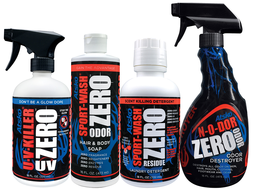
Important: First wash garment in Sport-Wash. Scent-Free Sport-Wash rinses 100% removing all traces of old soap residue, grease, oil, dirt, and sizing that would prevent U-V-Killer from forming a bond with the fabric. Other soaps do not rinse out and they leave behind residue. Sport-Wash is safe for all fabrics and dyes.

Spray Dry Garments. Spray till surface is covered. Imagine you are applying a color and don’t want the former color to show thru. Brushing with a small brush will make coverage more uniform. A ultraviolet (UV) light can reveal “missed” spots even while U-V-Killer is still wet. U-V-Killer extends the life of fabric five times from sun rot.

Fleeced or Insulated Garments. Extra care is required. You need to apply only a thin invisible coating. Spray a small area at a time. Use small stiff brush to raise froth on surface. Froth or foam bubbles keep the UV absorbing dye out on the visible surface while it dries. (This prevents the dye from soaking deep into the fabric where it will have no optical value). Deep soaking not only wastes U-V-Killer but will cause stiffness in heavy fabrics.

You may check the effectiveness and completeness of your application with a fluorescent black light. The bulb should appear dark VIOLET when not lit and will be labeled “BLB”. These are often used in bug zappers and we have a battery powered pocket unit available. Most paper contains UV brighteners and you can spray a spot to see how it will darken where U-V-Killer is applied.

All Fluorescent Orange has UV-Brighteners and most of it is water repellent. We have enhanced the wetting power of U-V-Killer for easier, more permanent application on Blaze Orange and other water repellent fabrics. You can be safe, legal, and successful.

Treated garments may be worn when completely dry, but 72 hours of adequate ventilation is required for the bond to reach full durability and for the garment to be scent free. Re-washing, waterproofing, and cover scent applications should be delayed for this 72 hours. The U-V-Killer treatment is designed to be permanent, and on most fabrics it will endure for the life of the garment.

A single washing in commercial detergents, fabric softeners, and or “Color Safe” bleach (while not removing the treatment) will render the U-V-Killer useless by depositing new brightener dyes on top of it.

Always wash a load of non-hunting clothes in Sport-Wash prior to washing your treated camo & blaze orange in order to remove residual brighteners from your washer and dryer. It is recommended that you confirm the absence of brighteners each season using a UV light.

FREQUENTLY ASKED QUESTIONS

  1. Will U-V-Killer, on my camo, cause skin irritation?
    No. U-V-Killer will not cause skin irritation unless someone is allergic to the dyes used on all clothing. When applying U-V-Killer , wash your hands when you are finished as there is no advantage to treating your skin.
  2. Will U-V-Killer cause my camo to fade or discolor?
    No. It will in fact protect your camo from fading caused by ultraviolet radiation. U-V-Killer is specifically formulated to be compatible with the dyes and fabrics used in the manufacturing of clothing. Poly/Cotton camo cloth tree blinds last 5 times as long if treated with U-V-Killer . It protects umbrellas, awnings, sail covers, tents and all outdoor fabrics from sun damage.
  3. What effect, if any, does U-V-Killer have on the cloth?
    With most fabrics there is no difference in feel, or hand as it is called in the textile industry; some fabric may feel slightly stiffer after drying but this slight stiffness disappears with wearing. If you want to eliminate this slight stiffness quickly before wearing, simply put the garment in your clothes dryer and tumble it with no heat for 15 minutes.
  4. Does U-V-Killer treat the inside of the garment when I spray it on the outside?
    No. You must spray the inside of lapels and pocket flaps to insure full treatment.
  5. How durable is the U-V-Killer treatment, and do I have to retreat my camo, and if so how often?
    On cotton garments U-V-Killer lasts forever, as long as you wash your camo in Sport-Wash. If you wash in Tide®, Cheer®, etc., you will have to retreat with U-V-Killer as you have redyed the cloth with brighteners. Synthetic fabrics last anywhere from 12 to 18 washings and then the garment must be retreated with U-V-Killer . So it is a good idea to retreat every season or two, especially knees, elbows and other areas of high abrasion. Normal wear (rain, snow, etc.) will not affect the U-V-Killer treatment.
  6. Can I treat my clothes with a waterproofing spray like Silicone Water-Guard after using U-V-Killer ?
    Yes. Waterproofing sprays work and have no optical effect on U-V-Killer . Always apply U-V-Killer first and allow to dry 72 hours before applying repellent. Be sure to test the water repellent for UV Brighteners. Many silicones and /or fluoropolymer repellents contain wax extenders that glow in UV. Those would defeat your purpose just as washing in the wrong detergent. Silicone Water-Guard Brand is free of brighteners as is our Permanent Water-Guard.
  7. How does U-V-Killer affect waterproof garments?
    Waterproof materials are still waterproof after treatment. Waterproof / Breathable membranes like Gore-Tex® are not affected at all. U-V-Killer does not cause any problems to seams, threads, or glue joints on any synthetic fabrics.
  8. I’ve treated my camo and I still see a blue glow with my UV light source. Does this mean that it is not working?
    Two possibilities exist. First, you may be seeing only visible blue and assuming it is UV. U-V-Killer does not block visible blue because blocking it would change the appearance of your camo in the visible spectrum, (We assume that you chose a pattern & color that appear to be right for the terrain you hunt). The UV light source, even the flashlight we sell, has some visible violet wavelengths present, and if all UV is absorbed you will still see some of this visible violet. If you place a single drop of U-V-Killer on a piece of brightened white paper or cloth and check it with the light source, you will see a dramatic difference because the brighteners are seen more on white. This demonstrates how effectively the absorber in U-V-Killer is working. You may be assured that a good application of U-V-Killer is just as effective on your camo and blaze orange.

    The second possibility is that the application wasn’t sufficient. Excess brighteners on the cloth will float to the surface of the U-V-Killer treatment. These excess brighteners must be removed by first washing with Sport-Wash. Wash twice if in doubt. Clean old detergent residue (which is full of brighteners) from washer by washing non-camouflage clothing inSport-Wash before washing your camo. Be sure to dry your camo (according to the care tag instructions) before treating with U-V-Killer . When applying U-V-Killer imagine you are applying a light coat of paint to the exterior surface. You want to cover every thread. If the spray is leaving spots, gaps, or streaks use a small brush, cloth, or even your hand to spread it back and forth. A bubbly white froth will remain visible up on the surface for a few minutes so that you can see that the coverage is complete. Check your progress with a black light.

    To properly check your camo under black light you must use a true Ultraviolet fluorescent light source. There are at least three domestic manufacturers of Ultraviolet tubes, for example- General Electric “BL”, Westinghouse “BL”, and Sylvania “Black light 350”. These black light tubes have radiation peaks at 350 nanometers. They will activate brighteners (like Tinopal by CIBA GEIGY) which are common in detergents. Manufacturers also offer “BLB” tubes. These are better because they have a blue filter to block visible wavelengths, they appear blue and are marked “BLB”. They cost more but make it much easier to detect UV Brighteners. A true UV light source will enable you to make a complete check of all your hunting gear and provides a way to double check yourself once you have treated your equipment with U-V-Killer products.
  9. Will U-V-Killer change the appearance of my camo? Will it look any different to me?
    No. All colors, camo, and blaze orange will look the same to you in full daylight.
  10. Will U-V-Killer crack or peel off at high or low temperatures?
    No. U-V-Killer forms a permanent bond.
  11. Is U-V-Killer flammable?
    No. In fact you can put out a fire with it.
  12. What about odor? Does U-V-Killer have any lingering scent?
    No. U-V-Killer is scent free when dry.
  13. Will U-V-Shield work on outdoor items that are exposed to severe weather?
    Yes. U-V-Shield is a clear weather proof coating that works great on items exposed to the elements such as duck decoys.
  14. Can I still spray my hunting clothes with a scent killer like N-O-DOR?
    Yes. U-V-Killer has no effect on N-O-DOR or how it works.
  15. Does Atsko make anything to make my Gore-Tex® jacket waterproof again?
    Yes. When your jacket was new it probably shed water because of a factory applied Fluoropolymer treatment on the outer shell. Contamination from wearing and residues of other detergents have made that repellent ineffective by providing pathways for water to wet through it. Wash the garment in Sport-Wash according to manufacturers recommendations. This will remove all residues and surfactants. Now iron the shell at the highest setting allowed by the manufacturer and the waterproofing will work like new again. If there was no factory waterproofing treatment, you can waterproof the garment with our Silicone Water-Guard. If the shell material is synthetic and can be ironed we recommend new Permanent Water-Guard water based fluoropolymer repellent. The results are guaranteed.
  16. What about use on Carbon Suits?
    All suits using carbon, charcoal, abscents, or any absorber or adsorber for odor control should be washed only in Sport-Wash. Treatment with U-V-Killer does not stop these garments from working. Remember other detergents leave residue which will clog up carbon suits and stop them from absorbing / adsorbing odor molecules.

Special Challenges for Application of U-V-Killer

There are several fabric structures that can interfere with the proper application of U-V-Killer and they can be extremely difficult to overcome. It is certainly preferable to select new camo without brighteners whenever possible.

One of the most common challenges is water repellent fabric. If you are applying the repellent, this problem is solved by simply applying the U-V-Killer first. After it is fully cured you can apply any non-glowing repellent right over it.

If the repellent is a silicone it can probably be removed in 2 or 3 washes in Sport-Wash. If it is factory applied, it will continue to repel U-V-Killer even after washing. You can increase the wetting power of U-V-Killer with the addition of I teaspoon of Sport-Wash or alcohol. It will also help to apply U-V-Killer right after the washer spin cycle instead of drying it first. It may also help to use a bit of fleece cloth or a veggie brush to spread U-V-Killer and work it into every cavity between fibers.

Mixed fibers like poly/cotton, while often no problem at all, can be near impossible to treat if the slicker fiber is the one that glows. The U-V-Killer treatment will drain into the more absorbent fiber leaving the glowing fiber untreated.

Absorbent fibers that have been surface treated can cause variable results. The problem is that printing, repellent, sizing, or other treatment may reduce the ability of the surface of the fiber to hold U-V-Killerwhile still allowing it to soak into the inside of the fiber. It may require several treatments to fill the fiber, and the surface may protect enough untreated brighteners to continue to glow. These items may look and feel like ordinary cotton but may not be treatable.

While many of these structures may accept treatment after many wearings and washings, when new, they may be impossible. If you treat only a small, hidden spot and it refuses to stay dead after several treatments you may be able to exchange the piece for something without brighteners. When all hunters have proper UV lights we suspect most marketers will increase their efforts to supply camo and blaze orange without brighteners.

Possible pretreatment like bleaching, abrasion, sealing etc. will certainly cause unacceptable damage, especially to a new garment. A thicker, heavy bodied product, like our U-V-Shield which is made for solid surfaces, could adhere better, but the increased thickness would stiffen the garment and make it abrasive and noisy. We have designed U-V-Killer to work on the widest possible range of fabrics but there are pieces that cannot be fixed. While all orange must be treated, you can save time, effort and money by selecting new camouflage that does not glow.

Special treatment for Difficult Items with U-V-Killer

With the help of feedback from some of the thousands of hunters who now have UV lights, we have identified difficult-to-treat items and special methods to treat them. Sometimes you can encounter a fabric that is not water repellent and appears initially to cover well, but when dry, it appears not to have been treated at all. This is not a failure of the absorbing dye, but rather a relocation of it to areas between, below, or inside the surface fibers where it can no longer block the uv energy that causes the brighteners to glow.

We have 2 techniques to overcome the problem which we have found successful on 100% polyester and 100% cotton. The first technique is presoaking. This apparently pre-fills with water, the voids that might otherwise become “black holes” for U-V-Killer to be lost in. Simply treat the item immediately after the final spin from the washer. The extra water may slightly dilute the U-V-Killer, but this is much better than having it disappear completely.

The low viscosity of U-V-Killer makes it spray and penetrate well, but also allows it to “creep” away before it dries. This can be minimized by applying heat immediately after treating. A small item like a hat could be done with a hair dryer, but for jackets, pants or coveralls, have your clothes dryer running on high temp and toss in each item as soon as you finish treating it.

These special techniques are not usually necessary, but they require no extra effort and testing the item with a spot of U-V-Killer will indicate if this extra attention is needed.

Behavioral Study Confirms That Deer See UV Brighteners

Bradley S. Cohen, David A. Osborn, George R. Gallagher, Robert J. Warren and Karl V. Miller


Although many aspects of white-tailed deer (Odocoileus virginianus) biology and physiology have been studied thoroughly, few studies have examined deer perception. We obtained a behavioral measure of light-adapted visual sensitivity based on performance of 7 deer in forced-choice discrimination tests. We compared intensity thresholds at the short and long wavelengths of their purported visual spectrum. Utilizing an automated training device, we trained deer to discriminate between lit and unlit light-emitting diode (LED) lights expressing monochromatic wavelengths, and recorded performance across a series of decreasing intensities for each wavelength until deer could no longer discriminate the light stimulus. We regressed a best-fit logistic function to each deer’s performance as intensity decreased at a single wavelength, demarcated the sensitivity threshold to that wavelength, and compared deer’s sensitivity threshold across wavelengths. Our behavior-based sensitivity measurements agreed with previous estimates of white-tailed deer’s cone photoreceptor sensitivity. We confirm that these cone photoreceptors contribute to deer behavior, and that deer have a greater perceptual sensitivity to shorter wavelengths and lower sensitivity to longer wavelengths. Our findings also suggest the deer have some sensitivity to ultraviolet light. The visual perception of the white-tailed deer is specialized for sensitivity during their crepuscular patterns, enables detection of predators along their horizon, and is well-adapted for a prey species. © 2014 The Wildlife Society.

Article first published online: 16 MAY 2014

DOI: 10.1002/wsb.438

Purchase article

Review of Color Vision in White-Tailed Deer (VerCauteren)

This is a comprehensive overview of the published studies of color vision in white-tailed deer. Published in December of 2003 it includes the owwos of those who preceeded Bradley Cohen’s team. This review by Kurt C. VerCauteren and Michael J. Pipas, both with the USDA in Fort Collins, CO enables you to trace the progress of over 60 years of work.

Cone and rod structures were identified in the deer retina in the 50s. By the 70s, pigments were isolated that could be shown to respond to colors of light. In August of 1993 Jerry Jacobs and Jay Neitz mearured the reponce of living deer eyes to different wavelengths of light. This enabled them to accuratley predict the spectral sensitivity of the deer.

All that remained was to design a behavioral experiment that could control for all other factors to prove that deer could see colors and ultraviolet by how they responded to different lights. Birgersson and others identified and resolved many of the issues that complicated this behavioral work. Then in February of 2012 at the 35th Annual South East Deer Study Group meeting Bradley Cohen’s work at the University of Georgia was publicized for the first time and it was published in the Wildlife Society Bulletin in May 2014.

Bradley Cohen’s study

VerCauteren and Pipas review